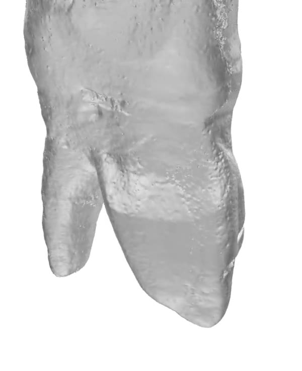
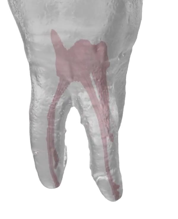
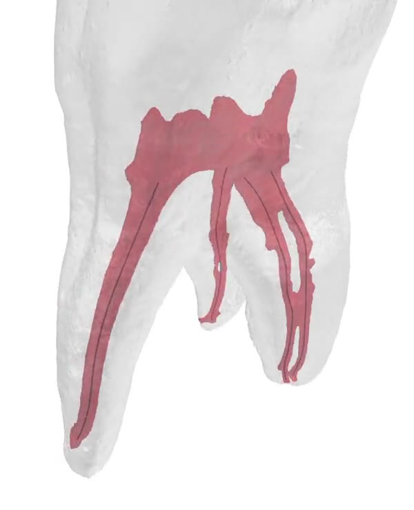
Maxillary first molar, specimen MX6.05. The root fades to show an H-shaped MB canal system: two canals, an isthmus between them, a re-split at the apex.
Read the canal,not the outline.
Every model is a scanned specimen, not an illustration.
Start with the real surface.
Micro-CT keeps every ridge and groove of the root. This is the tooth as it came out.
Fade the root.
Turn the surface translucent and the pulp shows through: the chamber, then each canal down to the apex.
Find the configuration.
MX6.05 has an H-shaped MB system. Two canals, joined by an isthmus, splitting again at the apex. You would not see it on a radiograph.
1Specimen MX6.05Maxillary first molar.
2Root surfaceSolid render from micro-CT.
1Pulp chamber
2CanalFollowed to the apex.
3Root, translucent
1MB canalTwo canals in one root.
2IsthmusJoins the two MB canals.
3Re-split at apexOne canal becomes two again.
MB2 loves to hide.
It sits under the dentin shelf, in a root you can’t see into. Study it on real specimens before you go looking for it with a file.
50+
Specimens toturn in your hand.
Canal networks, isthmuses, apical deltas, C-shaped canals, MB2 under the dentin shelf. All from real teeth.
Tag search
Find a configuration by name, for example “apical delta”.
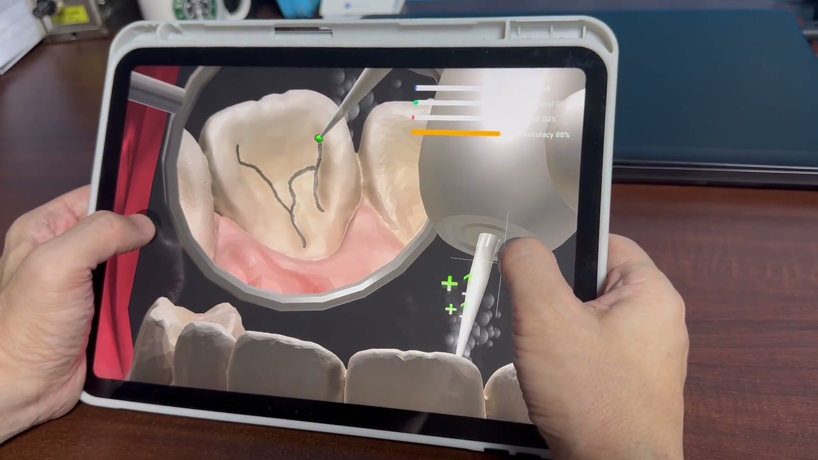
AR mode
Put the specimen in the room and walk around it.
Full-arch view
See each tooth where it sits in the arch.
Mastery sessions
Led by Seoul National University clinical instructors.
Smart Dentiform kit
Pairs with a physical kit, so the model in the app matches the one on your bench.
Watch the specimens.
Specimens turning, fading and splitting, posted at @dental_master_series.